
Figure 4.
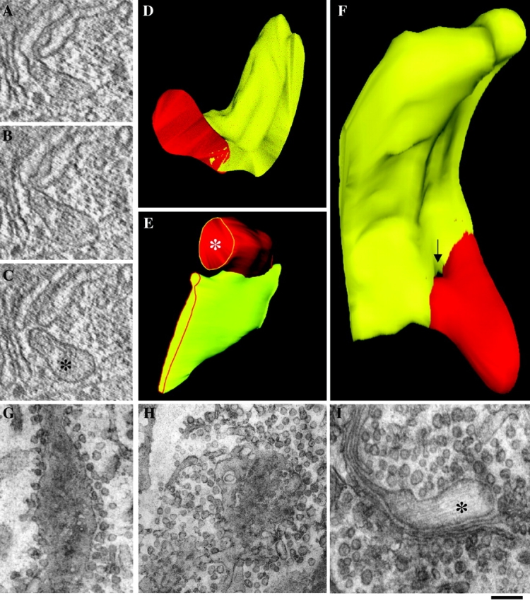
PC-I-containing distensions never dissociate from Golgi cisternae during intra-Golgi traffic. Human fibroblasts were subjected to the large ER accumulation–chase protocol (or to the large-pulse protocol) and fast frozen (A–F) or treated with NEM to inhibit vesicle fusion (G–I). (A–F) 9 min after the shift to 32°C, the cells were fixed by ultra-fast freezing, and then cryosubstituted and embedded into Epon 812. Thick (250 nm) sections of Golgi cisternae were cut, prepared for electron microscopic tomography, and virtual 2–3-nm slices (A–C) were extracted from the tomograms using the IMOD software (Ladinsky et al., 1999). The 3-D reconstruction and surface rendering of the cisternae (yellow) and distensions (red) were performed using the SURFdriver program. The same structure is shown in two orientations (D and F). The image in F was chosen to show a pore in the cisterna which generates the impression of discontinuity between distension and cisterna in one of the virtual sections (arrow). Serial thin (50 nm) sections of Golgi cisternae were cut and used to reconstruct the image in E. (G–I) NEM treatment. 7 min after releasing the temperature block, cells were placed on ice, and medium with (H and I) or without (G) NEM (100 μM) was added for 15 min. After washing on ice, cells were shifted to 40°C again for an additional 3 min, and then fixed and prepared for EM. Tangential section of a cisterna with surrounding vesicles in a control cell (G). Tangential section of a cisterna in a NEM-treated cell; vesicular profiles are much more numerous (three- to fourfold) than in controls (H). PC-I distension connected with a cisternae in a NEM-treated cell (I). *, PC-I–containing distensions. Bar: (A and C–E) 150 nm; (F) 100 nm; (G and H) 300 nm; (I) 200 nm.