
Figure 3.
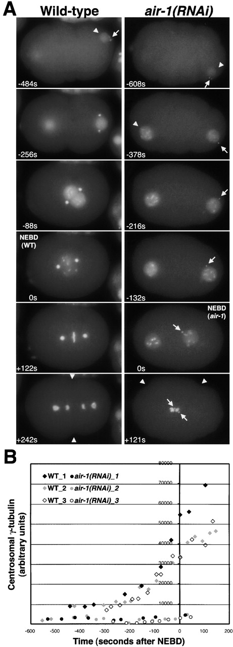
AIR-1 is required for the accumulation of centrosomal γ-tubulin during maturation. (A) Panels summarize time-lapse recordings of wild-type (left) and air-1(RNAi) embryos (right) expressing GFP histone and GFP–γ-tubulin. Both sequences start at a similar early embryonic stage, judged by the extent of cortical ruffling, the small size of the sperm pronuclei (arrowheads), and the lack of separation of γ-tubulin–labeled centrosomes (arrow). The sequences end with onset of cytokinesis (wild-type, +242 s) or cortical contractions (air-1(RNAi), +121 s, arrowheads). The duration of both recordings is ∼12 min. Times are seconds after NEBD. The times in the corresponding panels differ by ∼2 min because NEBD is slightly delayed in air-1(RNAi) embryos. See videos 3–6 available at http://www.jcb.org/cgi/content/full/jcb.200108051/DC1. (Left) In wild-type, the DNA condenses, beginning before the migration of the pronuclei toward each other and continuing during migration. This occurs concomitant with an increase in the amount of centrosomal γ-tubulin (compare −256 s with −88 s). The accumulation of centrosomal γ-tubulin continues after NEBD as the mitotic spindle assembles (+122 s). (Right) In air-1(RNAi) embryos, the chromosomes condense with timing similar to wild-type, but γ-tubulin fails to accumulate at centrosomes (arrows, all panels). The migration of the maternal pronucleus toward the sperm pronucleus is lethargic, probably due to the failure of centrosomes to nucleate robust mitotic asters, but the two pronuclei eventually move toward each other, and the nuclear envelope breaks down (0 s). Chromosomes never align properly, but cortical contractions begin coincident with cytokinesis in wild-type embryos (+121 s). Cytokinesis does not succeed in air-1(RNAi) embryos (unpublished data). (B) Kinetic traces of centrosomal γ-tubulin fluorescence. Centrosomal fluorescence was quantified in 7 wild-type and 10 air-1(RNAi) embryos. Three traces are shown for each. Bar, 10 μm (as in Fig. 2).