
Figure 4.


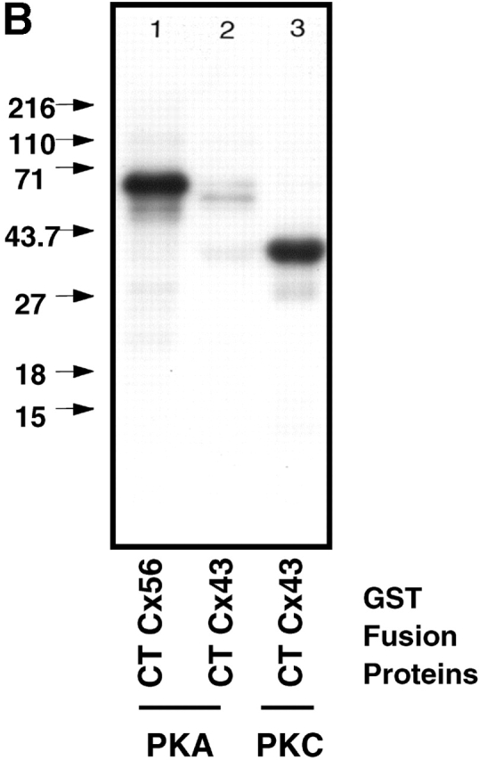
S364 of Cx43 is phosphorylated in unstimulated WT Cx43/KO fibroblasts. (A) HPLC elution profile for 32P-labeled Cx43 peptides isolated from cells expressing WTCx43 (•) or S364PCx43 (□). The elution position of certain peptides is indicated. Amino acids 346–382 of Cx43 and tryptic cleavage sites are depicted above the profile (arrows). Asterisks denote sites that may only be partially cleaved by trypsin. (B) Autoradiogram depicting in vitro phosphorylation of Cx GST fusion proteins. (Lane 1) Purified GST CTCx56 phosphorylated by PKA. (Lane 2) Purified GST CTCx43 phosphorylated by of PKA. (Lane 3) Purified GST CTCx43 phosphorylated by PKC. (C) Phosphorylation of Cx43 after treatment with 8Br-cAMP. Autoradiogram showing results of two separate experiments in which confluent monolayers of WT fibroblasts (10-3) were loaded with radiolabeled [32P]i in the presence or absence of 8Br-cAMP (Materials and methods). (1 and 3) Untreated. (2 and 4) Treated with 8Br-cAMP.