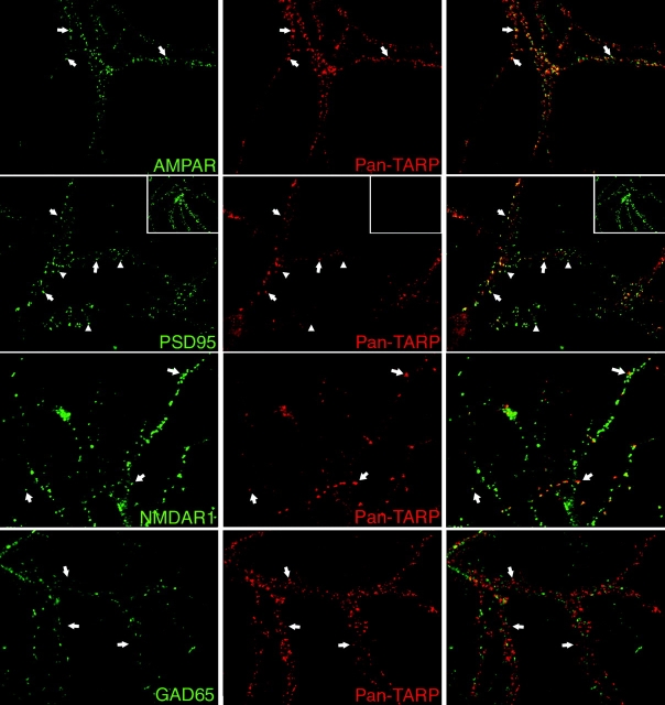
Figure 5.
TARPs cluster selectively at excitatory synapses that contain AMPA receptors. An antibody (pan-TARP) that reacts with all TARPs was used to stain cultured hippocampal cultures. (First row) Immunofluorescence for TARPs occurs at punctate sites (arrows) along the dendrites that closely colocalize with the GluR2 subunit of the AMPA receptor (AMPAR). (Second row) Virtually all TARP puncta colocalize with PSD-95 (arrows), but some PSD-95 puncta lack TARP immunofluorescence (arrowheads). (Third row) Almost all TARP puncta colocalize with NMDAR1 (arrows), but some NMDAR1 puncta lack TARP immunofluorescence. (Fourth row) TARPs show no overlap with the GABAergic marker GAD65. Preabsorbing the antibody with antigen (10 μM) blocks labeling (small boxes in second row).

