
Figure 2.
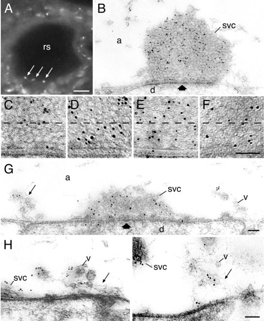
Redistribution of synapsin in resting and stimulated reticulospinal synapses. (A) Transverse section of a reticulospinal axon (rs) stained with lamprey-specific synapsin antibodies using immunofluorescence. Punctate labeling (arrows) is localized close to the surface of the axon. (B) The ultrastructural localization of synapsin at rest. Gold particles are associated with the synaptic vesicle cluster. Thick arrows indicate active zone. (C–F) An increase in synapsin labeling in the area within 100 nm from the presynaptic membrane in the vesicle cluster at different states: (C) rest; (D) 5 Hz action potential stimulation; (E) high K+ stimulation; (F) control, SV2 immunolabeling at rest. (G–I) Electron micrographs of synapses stimulated by action potentials at 5 Hz for 20 min and labeled for synapsin. Thin arrow indicates filamentous matrix. Note that synapsin is associated with the filamentous cytomatrix and vesicles in the endocytic zone. svc, synaptic vesicle cluster; v, vesicles in the filamentous matrix; d, dendrite; a, axoplasmic matrix. Bars: (A) 20 μm; (F) 100 nm, also for C–E; (G) 100 nm, also for B; (I) 100 nm, also for H.