Figure 4.
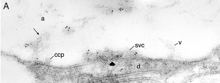
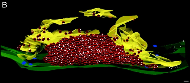
3-D distribution of synapsin in stimulated reticulospinal synapse. (A) Electron micrograph of one serial section of a reticulospinal synapse stimulated at 5 Hz for 20 min, shown reconstructed in B. Synapsin is present on vesicles (v) in the filamentous cytomatrix (arrow). svc, synaptic vesicle cluster; v, vesicles in the filamentous matrix; d, dendrite; a, axoplasmic matrix. ccp, clathrin-coated pit. This arrow indicates active zone. (B) 3-D reconstruction of 10 serial ultrathin sections. The area occupied by actin-rich filamentous cytomatrix lateral to the vesicle cluster is shown in yellow. The plasma membrane is depicted in green. All synaptic vesicles are shown in red. Colloidal gold particles are indicated by white spheres. Clathrin-coated endocytic intermediates are shown as flat blue discs on the plasma membrane. Bar, 100 nm.


