
Figure 3.
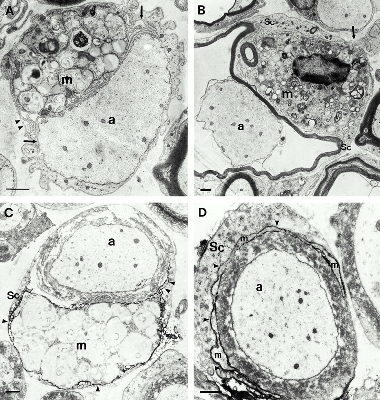
Conventional electron microscopy (A and B) and immunoelectron microscopy using the macrophage-specific antibody F4/80 (C and D) in quadriceps nerves of 6-mo-old P0+/− mice. (A) A putative macrophage (m) that is laden with myelin debris is closely apposed to a demyelinated axon (a). Note the slender processes of the macrophage (arrows) and the position of the cell within the endoneurial tube. The arrowheads demarcate the Schwann cell basal lamina. (B) A putative macrophage (m) is in close contact to a thin myelin sheath that is partially detached from the corresponding axon (a). The arrow demarcates a process of the putative macrophage that penetrates the Schwann cell basal lamina. Sc, Schwann cell. (C) An F4/80-positive macrophage (m) containing myelin debris is in close apposition to a myelin sheath. Arrowheads indicate electron-dense immunoreaction product. a, Axon; Sc, Schwann cell. (D) A slender process of a F4/80-positive macrophage (m) has penetrated in between the pericaryon of the Schwann cell (Sc) and its normal appearing myelin sheath. Arrowheads indicate electron-dense immunoreaction product. a, axon. Bars, 1.5 μm.