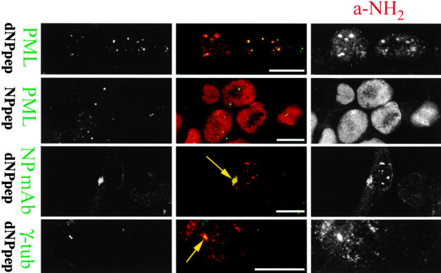
Figure 3.
dNPpep rescued by zLLL localizes in PODs and the MTOC. rVV-infected 143B cells were incubated for 3 h before the addition of 20 μM zLLL. After 6.5 h, cells were fixed, permeabilized, stained with the indicated antibodies, and imaged with the LCSM. The second row displays cells expressing NPpep, in the first and third, cells expressing dNPpep. In the bottom row, dNPpep expressing cells were first extracted with 1% NP-40 and then fixed with methanol/acetone (80:20) for 15 min at −20°C to enable staining with the γ-tubulin–specific mAb. Arrows point to the MTOC. Gray-scale images on the sides are merged in the middle with the color indicated by the text describing the antibody specificity. Bar, 10 μM.

