
Figure 7.
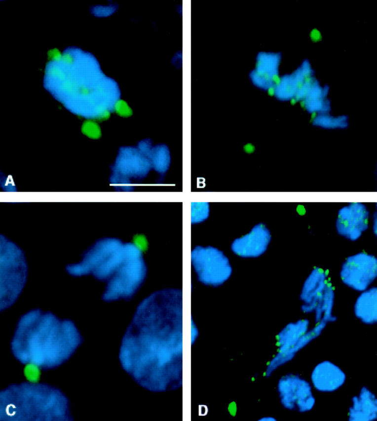
Distribution of 3F3/2 phosphoepitopes in bub1 mutant neuroblasts. DNA is in blue, and 3F3/2 phosphoepitopes are in green. In all panels, the two strongest sites of 3F3/2 staining are the centrosomes. 3F3/2 distribution in prophase (A) and metaphase (B) neuroblasts from bub1 mutants. 3F3/2 epitopes at the centrosomes and kinetochores are strongly recognized, demonstrating that the Bub1 kinase is not a significant source of 3F3/2 phosphorylation activity in vivo. (C) 3F3/2 epitopes are completely dephosphorylated during anaphase in wild-type neuroblasts (see Bousbaa et al. 1997 for a detailed description of 3F3/2 distribution in wild-type Drosophila neuroblasts). (D) 3F3/2 distribution in an anaphase figure from a bub1 mutant neuroblast. 3F3/2 epitopes continue to remain phosphorylated in bub1 anaphases, though at reduced levels relative to those seen during prophase/prometaphase. Thus, total dephosphorylation of 3F3/2 phosphoepitopes cannot be a prerequisite for entry into anaphase. Bar, 5 μm.