Figure 6.
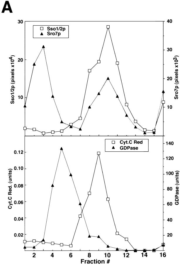
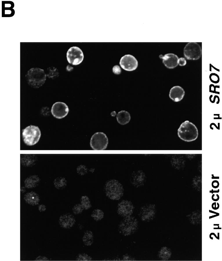
Sro7p is associated with the plasma membrane in yeast. (A) Sro7p colocalizes with the plasma membrane marker Sso1/2p on a sucrose density gradient. Wild-type cells grown to logarithmic phase were spheroplasted, lysed, and spun at 30,000 g to pellet the membrane-associated pool of Sro7p that was analyzed on a 20–50% sucrose density gradient. The top shows the fractionation profile of Sro7p and the plasma membrane t-SNARE Sso1/2p in the gradient as determined by immunoblotting. The bottom shows the distribution of the GDPase activity (a Golgi marker) and cytochrome c reductase activity (an ER marker) in the gradient. Fraction 1 corresponds to the top of the gradient. Sro7p is found in two pools on the gradient: one that has been released from the membrane and is found at the top of gradient and a second pool that comigrates with the plasma membrane marker Sso1/2p. (B) Indirect immunofluorescence localization of Sro7p to the plasma membrane and soluble pool of the cell. Wild-type cells containing vector only (pB23) or SRO7 on high copy (pB497) were grown in selective media overnight and shifted in YPD for 2 h before fixing with formaldehyde. Cells were permeabilized using 0.05% SDS and stained with affinity-purified α-Sro7p antibody and detected using a rhodamine-conjugated α-rabbit secondary antibody. Stained cells were observed on a Zeiss LSM 510 confocal microscope and individual Z-slices were captured with LSM 510 software. Plasma membrane and cytosolic staining is evident in cells containing the high copy SRO7 but no specific staining was observed in the empty vector control cells containing a single copy of SRO7.


