
Figure 2.
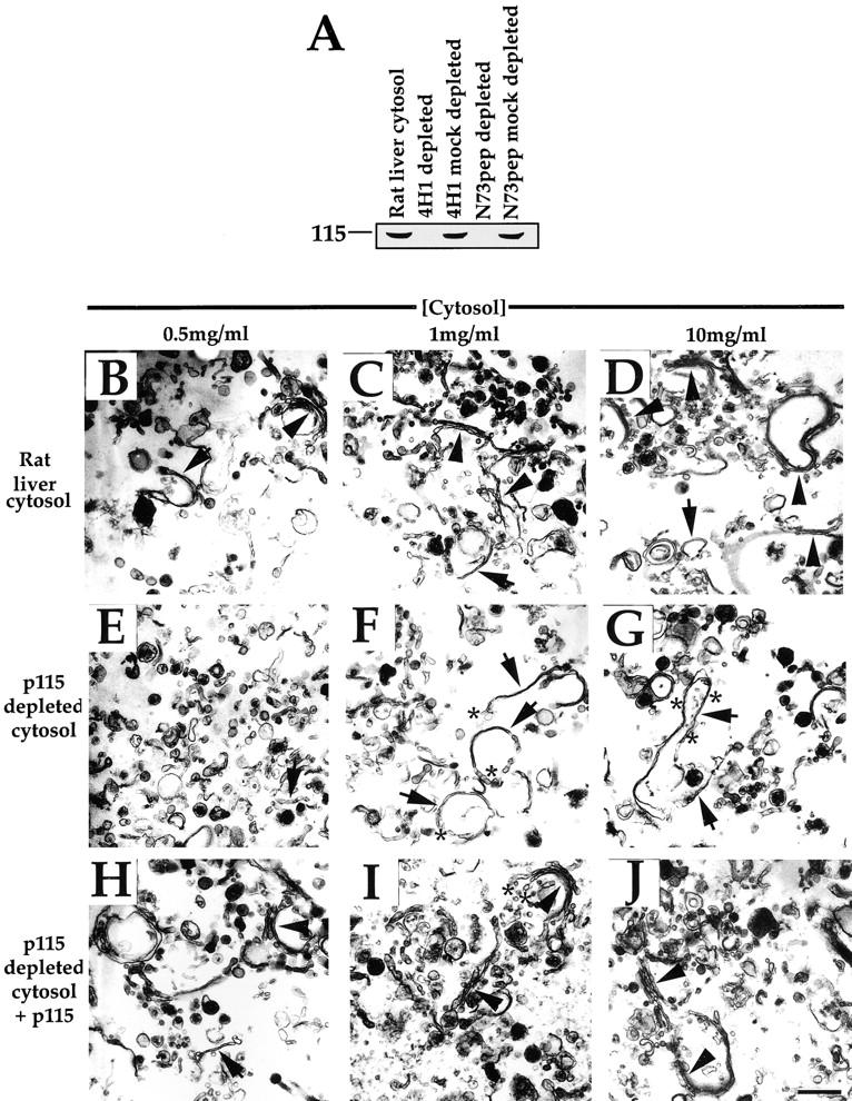
Effect of interphase cytosol depleted of p115 on the reassembly process. (A) Rat liver cytosol was depleted of p115, as described in Materials and Methods, using either the anti-p115 mAb antibody 4H1 or the N73pep. 20 μg of cytosol was fractionated by SDS-PAGE using a 7.5% gel, transferred to nitrocellulose, and probed with the anti-p115 mAb 8A6. (B–J) MGF isolated through a 0.5-M sucrose cushion were incubated for 60 min at 37°C with increasing concentrations of either rat liver cytosol (B–D), p115-depleted cytosol (E–G), or p115-depleted cytosol supplemented with purified rat liver p115 (H–J), fixed, and processed for EM. Representative fields are shown. Note the presence of stacks (arrowheads) in B–D and H–J, but only single cisternae (arrows) in E–G. The reduced number of cisternae in E suggests poor reassembly at this concentration of p115-depleted cytosol. Note that the cisternae formed often have a wrinkled appearance (asterisks in G) in p115-depleted cytosol and are often blunt-ended with few associated vesicles (compare asterisks in F and I), in contrast to when p115 is present. Bar, 0.5 μm.