Figure 2.
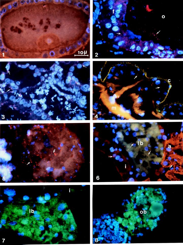
FISH of Wolbachia (red) and SOPE (green) in different weevil tissues. (1 and 2) Comparison between the Wolbachia-negative control (1) [oocyte from the SOPE-aposymbiotic Chinese strain (ChH) treated with tetracycline (ChHT)] and the Wolbachia-positive control (2) [oocyte (o) and follicular cells (f) of the SOPE-aposymbiotic strain (ChH)]. (3) Testes of the SOPE- and Wolbachia-symbiotic strain (Ch) (see arrows, Wolbachia near spermatid nuclei). (4) Cuticle (c) and muscle (m) of the SOPE- and Wolbachia-symbiotic strain (Ch). (5) SOPE-aposymbiotic apex of ovary (ChH). (6) Intestine (i), the larval bacteriome (lb) in green and the adipocytes (a) of the Ch strain: both SOPE (green) and Wolbachia (red spots, arrows) are visible in the bacteriocytes. (7 and 8) Larval bacteriome (lb) and the ovarian bacteriome (ob) of the French Wolbachia-free strain (SFr). All slides were hybridized with the three probes (W1, W2, and S; see Materials and Methods). (Scale bar, 10 μm.)

