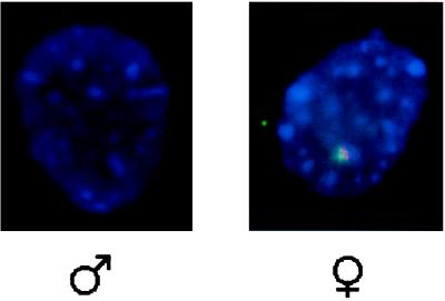
Figure 4.
RNA–FISH photomicrograph of male and female somatic cells. RNA–FISH was performed to visualize cytoplasmic/nuclear RNA that hybridized to Xist probes pWS850 (FITC) and pWS854 (rhodamine). Hybridizations of Xist probes were performed simultaneously and separate channels recorded; they were merged after recording. Micrograph shows the colocalization of the probes pWS850 and pWS854. (×600.)

