
Fig. 4.
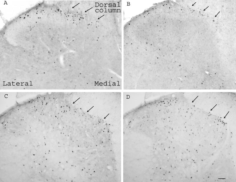
Microphotographs showing Fos expression in the spinal cord in rats with sham operation (A, B) or dorsolateral funiculus lesion (C, D). Note that EA (B) inhibited Fos expression compared to sham EA (A) in sham-operated rats, and that EA (D) did not show significant inhibition of Fos expression compared to sham EA (C) in rats with dorsolateral funiculus lesioning. Arrows point to medial superficial laminae. Scale bars are 50 μm.