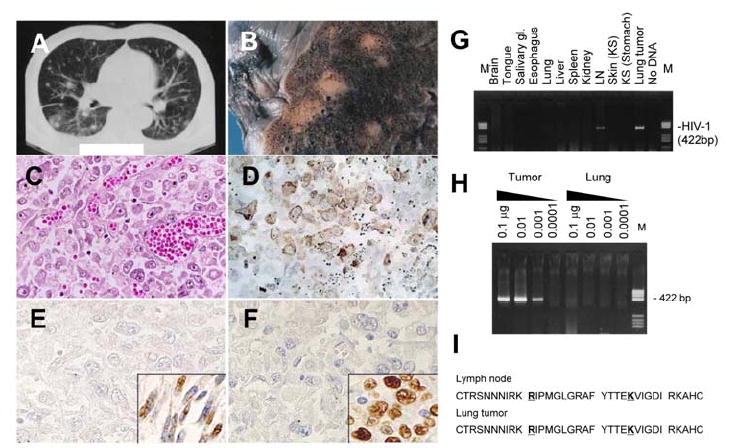
Figure 1. Pathological findings of tumors in the lung of a patient with AIDS.

CT scan (A), macroscopic view (B) and Hematoxylin and eosin staining (C) of the lung tumor. (D) Immunohistochemistry of CD45RO. (E) Immunohistochemistry for KSHV-LANA in the lung tumor cells. Inset shows gastric KS cells from the patient. (F) In situ hybridization for EBV-EBER in the lung tumor cells. Inset shows a positive control of EBV-positive lymphoma from an unrelated patient. (G) PCR detection for HIV-1 V3 region in various organs of the patient. LN, lymph node; M, DNA molecular weight marker (pBR322/HaeIII). (H) Semi-quantitative PCR for HIV-1. DNA quantities are indicated at the top of the panel. DNA extracted from the lung tumor and surrounding lung tissues was tested. (I) Predicted amino acid sequence of HIV-1 gp120 V3 loop of HIV-1 amplified from the lymph node and lung tumor by PCR. Positions 11 and 25 are indicated by bold letters with underlines. DNA sequences are deposited in GenBank under accession numbers DQ116951 to DQ116954 (HIV-1 envelope from LN and lung tumor).
