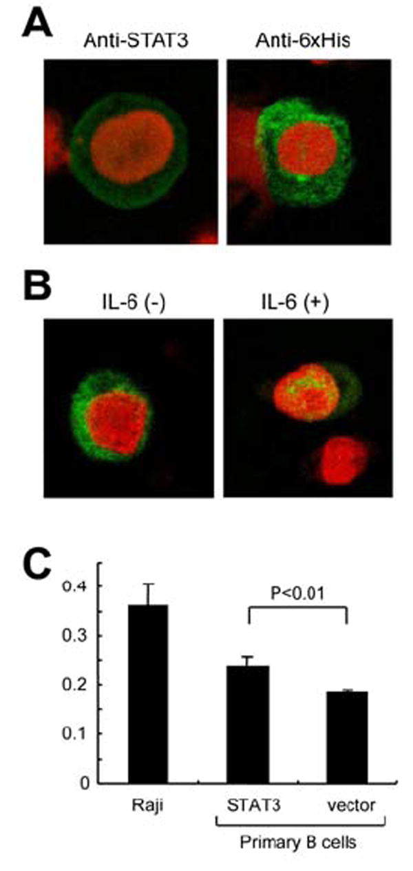
Figure 5. Transfection of STAT3 into B cells in vitro.

(A) STAT3 expression in the STAT3-transfected TY-1, a KSHV-positive B cell line. The cells were transfected with STAT3 expression vector by Nucleofector (Amaxa, Cologne, Germany) using O-06 program. STAT3 expression was detected by anti-STAT3 mouse monoclonal antibody (green in left panel) and anti-6xHis antibody, followed by Alexa 488-conjugated anti-mouse IgG antibody (Molecular probe, green in right panel). Red color indicates nuclear counterstaining of propidium iodide. (B) Localization of transfected STAT3 in TY-1. His-tagged STAT3 was detected by anti-6x His antibody in the cytoplasm of B cells (left panel). In the presence of IL-6 (Peprotech, Rocky Hill, NJ, 0.1 ng/ml), transfected STAT3 localizes in the nucleus predominantly (right panel). (C) Cell proliferation assay for STAT3-transfected primary B lymphocytes. Primary B cells were isolated from PBMC. The purity of B cell (CD19+) was >95%. The cells were transfected with STAT3 expression vector expressing STAT3 and CD4 by Nucleofector using U-15 program. Transfection efficiency to primary B cells was around 20%. To increase the proportion of transfected cells, the transfected B cells were separated with CD4 microbeads after 16 hours of the transfection (Miltenyl Biotec, Auburn CA). 48 hours after transfection of STAT3 or vector to primary B cells, the proliferation rate was measured with BrdU ELISA (Roche). Raji is an EBV-positive Burkitt lymphoma cell line (untransfected). Numbers in Y-axis indicates absorbance in ELISA. Error bars indicate standard errors of 8 independent experiments.
