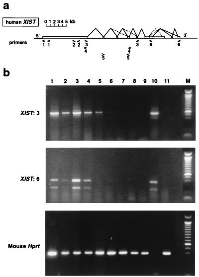
Figure 2.
RT-PCR analysis of human XIST expression in transgenic mouse ES cells. (a) The human XIST gene is shown with the locations and orientations of the six primer pairs (1–6) used (see Materials and Methods). The previously determined splicing pattern (7) is shown, with less frequently observed splicing events indicated by dashed lines. (b) Amplification of cDNA in transgenic cells before and after differentiation (8-day EBs). Positive control human cDNA is female brain. Mouse controls are the host ES cell line CK35 and female kidney. Lanes: 1, hY20 undifferentiated; 2, hY20 EBs; 3, hY21 undifferentiated; 4, hY21 EBs; 5, hY1 undifferentiated; 6, hY1 EBs; 7, hY5 undifferentiated; 8, hY5 EBs; 9, CK35 undifferentiated; 10, human female brain; 11, mouse female kidney. Representative data using primer pairs 3 and 6 are outlined in a. Mouse Hprt RT-PCR was used to control the RT samples. The PCR products shown are cDNA-specific as the primers flank introns.

