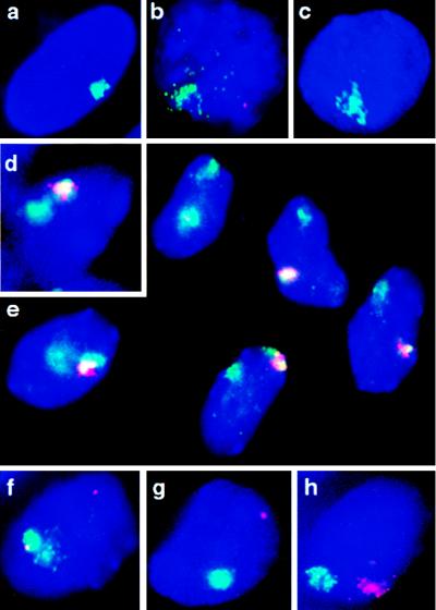
Figure 3.
FISH analysis of human and murine Xist expression in transgenic mouse ES cells. RNA FISH on undenatured nuclei and DNA FISH after chromatin denaturation (see Materials and Methods). Nuclei are counterstained with 4′,6-diamidino-2-phenylindole (blue). (a) Human XIST RNA (green) detected in human amniocytes: a domain-like signal over the human X chromosome (8). (b) Representative example of human XIST RNA domain (green) and mouse Xist RNA pinpoint (red) signals in undifferentiated ES cells of line hY20 or hY21. (c) Human XIST RNA domain (green) in line hY21 after differentiation (8 days). (d) Human XIST RNA (red) and chromosome 2 DNA (green) in transgenic hY20 cells; overlapping signals appear yellow. (e) Multiple differentiated cells of transgenic line hY21, illustrating the degree of localization of the human XIST RNA (red) over chromosome 13 (green), which was high in the majority of cells and more restricted (e.g., top left) or dispersed (e.g., bottom left) in a minority of cells. Similar observations were made in undifferentiated hY21 cells. Overlapping signals appear yellow. (f) Human XIST RNA (green) and Dhfr/Rep-3 RNA (red) signals detected in undifferentiated hY21 nuclei. (g) Human XIST RNA (green) and Dhfr/Rep-3 RNA (red) signals detected in differentiated (10-day) hY21 cells. (h) Human XIST (red) and mouse Xist (green) RNA domains detected in differentiated (10-day) transgenic hY21 cells.

