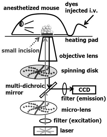
Figure 1. Schematic diagram of the confocal microscope for real-time in vivo imaging.
To visualize cell dynamics in vivo, fluorescent dyes were injected into the tail veins of anesthetized mice. A small incision was then made for an observation window, and the mouse was set on a heating pad to maintain body temperature. An inverted microscope equipped for Nipkow–spinning disk confocal laser microscopy, which enabled scanning at up to 1,000 frames/s, was used to visualize the tissue. See Methods for details.

