
Figure 7. Wnt Signaling Induces Epidermis in a Smad1/5/8 and β–Catenin Dependent, but Tcf3 Independent, Manner.
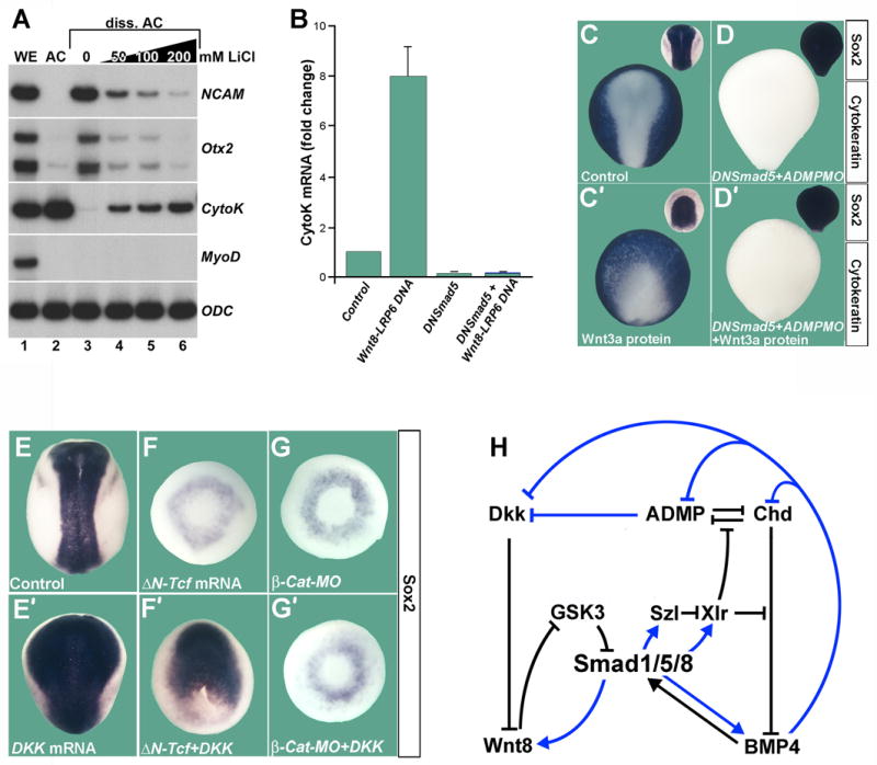
(A) LiCl induces epidermis (Cytokeratin) and inhibits neural differentiation (NCAM, Otx2). Radioactive RT-PCR analysis of whole embryos (WE), animal cap (AC) explants and dissociated animal cap cells at stage 13. MyoD indicates lack of mesoderm induction and Ornithine decarboxylase (ODC) equal loading.
(B) Quantitative PCR of dissociated animal caps injected with pCSKA-Wnt8 and pCS2-LRP6 DNA. DN-Smad5 mRNA was co-injected to block Smad1/5/8 activity. Cytokeratin mRNA levels at stage 13 were normalized for ODC mRNA and the standard deviation from three independent experiments is indicated.
(C,C′) Wnt3a protein (60 nl of 16 ng/μl) microinjected into the blastula cavity at stage 9 inhibits anterior neural plate and expands epidermis (n=40 and 42, respectively).
(D,D′) DN-Smad5 converts the entire ectoderm into neural tissue and is epistatic to Wnt3a protein injection (n=27 and 35). ADMP MO was co-injected to eliminate all traces of epidermis. (E,E′) Dkk1 mRNA expands the neural plate (n=100 and 53).
(F) ΔN-Tcf3 mRNA eliminates the neural plate; only a ring of Sox2 expression in ventral mesoderm remained (90%, n=30).
(F′) Dkk1 mRNA rescues neural plate in the presence of ΔN-Tcf3 in 60% of embryos (n=70).
(G,G′) The induction of neural plate by Dkk1 mRNA has a complete requirement for β-Catenin (100%, n=17 and 55, respectively).
(H) Model in which the BMP (D–V) and Wnt (A–P) patterning pathways are integrated at the level of Smad1/5/8 phosphorylations. Black arrows indicate direct protein-protein interactions and blue arrows transcriptional regulation by Smad1/5/8; all interactions are supported by overexpression or morpholino studies in Xenopus (Lee et al., 2006, and data not shown).