
Figure 1.
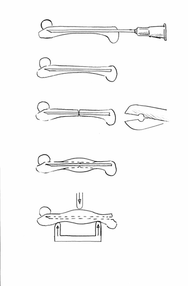
Overview of experimental set-up. After insertion of the intramedullary cannula,, the specially made pair of scissors with semi-lunar cutting edges was slid along the bone to approximately mid-diaphysis and the femur cut.
Official websites use .gov
A
.gov website belongs to an official
government organization in the United States.
Secure .gov websites use HTTPS
A lock (
) or https:// means you've safely
connected to the .gov website. Share sensitive
information only on official, secure websites.
Overview of experimental set-up. After insertion of the intramedullary cannula,, the specially made pair of scissors with semi-lunar cutting edges was slid along the bone to approximately mid-diaphysis and the femur cut.