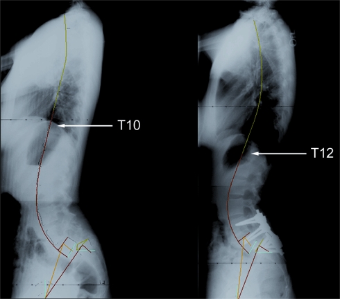
Fig. 3.
Pre- and post-operative lateral radiographs of a patient with developmental spondylolisthesis. Pre-operatively, the patient has a kyphotic segment that spans from T1 to T10 and a lordotic segment from T10 to S1. Post-operatively, the kyphosis is from T1 to T12 and the lordosis is from T12 to S1

