
Fig. 6.
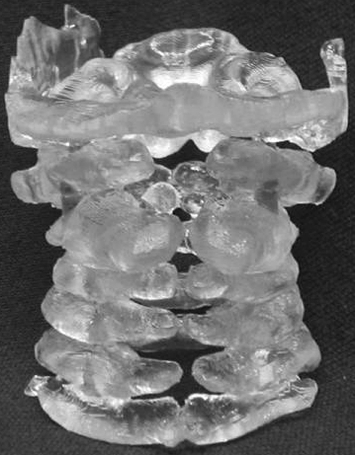
Posterior view of biomodel of cervical spine (SEDC in Case 20) illustrating the posterior bony deficits, which were not clearly demonstrated by other investigations. The biomodel allowed safer exposure of the spinal cord and facilitated the placement of posterior instrumentation to achieve fusion