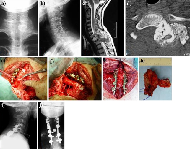
Fig. 1.
Patient no. 3. Extracompartmental osteosarcoma affecting the Th1 vertebra in a male 12-year-old child, symptomatic with acute paraparesis and necessitating emergency laminectomy. Front (a) and lateral (b) radiographs show a large radiodense tumor mass extending dorsal and left to Th1. Sagittal MRI scan prior to laminectomy (c) revealed complete destruction of the dorsal elements, spinal canal involvement and marked compression of the spinal cord. Following emergency laminectomy and neoadjuvant multimodal therapy (COSS 96 and radiation), coronar CT-scans (d) demonstrate a decrease in size and increase in sclerosis of the tumor tissue. (e) Intraoperative view showing rotation of the Th1 vertebra around the longitudinal axis. After en bloc excision (f) posterior cervicothoracal reconstruction with anterior expandable cage interposition (g) was performed. Specimen (h) of Th1-vertebral osteosarcoma originating from and extending dorsolaterally to the pedicle after en bloc excision, showing histologically tumor-free margins (posterior view). (i), (j) Postoperative biplanar radiographs following dorsoventral instrumentation

