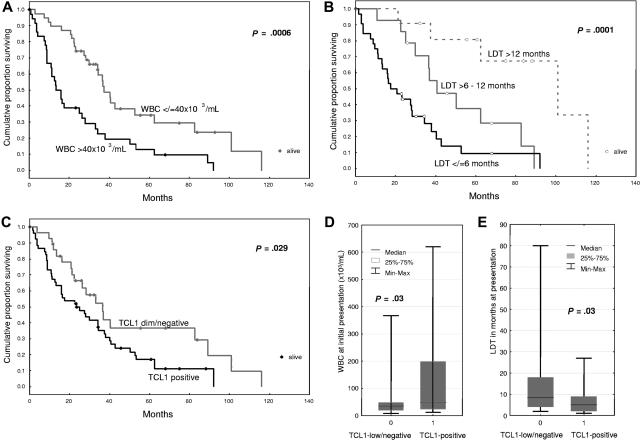
Figure 1.
The molecular feature of TCL1 expression is associated with a hyperproliferative clinical subgroup of T-PLL. Kaplan-Meier analysis of overall survival in T-PLL patients showed an inverse association with presenting white blood cell (WBC) count (A), pretreatment lymphocyte doubling time (LDT, B), and TCL1 immunohistochemical expression (C). The TCL1+ subset of T-PLL showed higher presenting WBC counts (D) and shorter pretreatment LDT (E).

