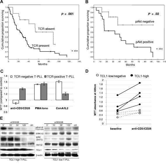
Figure 2.
The T-cell receptor signaling pathway is functional in T-PLL and responses are influenced by TCL1 levels. (A) Detection of surface T-cell receptor (sTCR) in T-PLL cells by flow cytometry for sCD3 and TCRα/β (67/82 sTCR+ versus 15/82 sTCR− cases) correlated with shorter overall survival (OS). (B) Expression of the Ser473-phosphorylated activated form of AKT, as analyzed by Western blot, also significantly correlated with poor OS. (C) TCR engagement stimulated growth in T-PLL tumor cells only in cases expressing sTCR, whereas ConA/IL2 or PMA/ionomycin stimulated growth in nearly all cases (MTT assay values normalized to unstimulated control). (D) In sTCR+ T-PLL, the degree of growth induction by TCR engagement (MTT assay) was higher in those tumors that strongly expressed TCL1 (solid dots) compared with TCL1-low/negative tumors (white circles). (E) Western blot analysis reveals that T-PLL with higher TCL1 levels was associated with a faster induction of pERK1/2 and activated AKT (left versus right panels).

