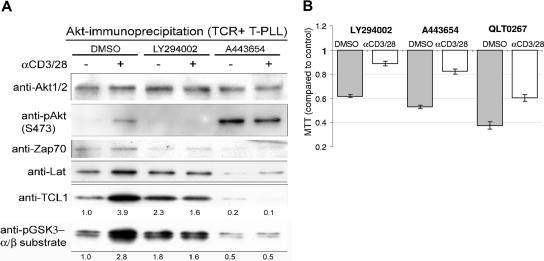
Figure 4.
Inhibitors of AKT signaling alter AKT-TCL1 complex formation and T-PLL growth in culture. (A) AKT complexes were isolated from primary T-PLL lysates by immunoprecipitation using an immobilized anti-AKT antibody, and showed increased phosphoactivated (pS473) AKT and TCL1 as well as increased amounts of ZAP70 and LAT following 30 minutes of TCR engagement. Increased AKT kinase activity following TCR stimulation was found by elevated phospho-GSK3α/β substrate levels (numbers indicate fold changes following stimulation compared with DMSO control). Preincubation of T-PLL tumor cells with the PI3K inhibitor LY294002 or the AKT inhibitor A443654 abolished the TCR-stimulated phosphorylation of AKT and GSK3α/β target and decreased the amount of TCL1 complexed with immunoprecipitated AKT. A443654 also led to an overall increase in pAKT-S473. (B) LY294002 at 20 μM, A443654 at 0.5μM, and QLT0267 at 40 μM reduced the level of proliferation in unstimulated T-PLL cultures at baseline and following TCR cross-linking compared with the DMSO vehicle control (MTT assay at 48 hours).

