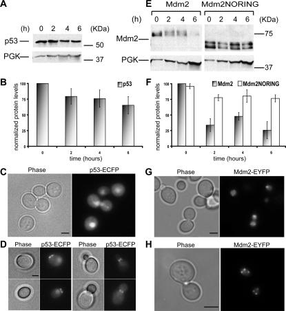
Figure 1. Stability and localization of the human p53 and Mdm2 proteins in yeast.
(A) Total yeast cell extracts from exponentially growing cells were immunoblotted for p53 over time. 3-phosphoglycerate kinase (PGK) was used as loading control. (B) Quantifications of p53 protein levels are shown. Data represent the mean ± standard error (SE), for n = 3 independent experiments. (C,D) Maximum projections from fluorescence image stacks of live yeast cells expressing p53-ECFP were obtained with a DeltaVision workstation. Bar, 2 µm. E,F,G,H. Same as in (A,B,C,D) but for Mdm2.

