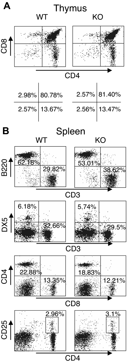
Figure 3.
Normal T- and B-cell development in thymus and spleen of Rsk2 KO mice. Representative flow cytometric profiles of thymus (A) and spleen (B) from WT littermate and Rsk2-deficient mice (KO). The numbers show the percentage of each cell population with positive staining by indicated antibodies.

