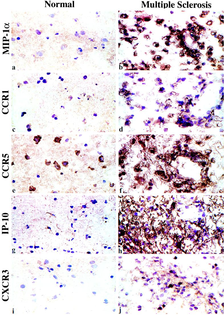
Figure 3.
Immunopathology of progressive MS illustrating chemokine and chemokine receptor expression within the CNS in an area of demyelination (Right). Left micrographs show the very limited expression of these proteins in normal brain, with focal expression of CCR5 by neurons, and IP-10 expression by astrocytes; only minor expression, comparable to normal brain, was seen in the uninvolved tissues of MS patients with inactive disease (not shown). In contrast, areas of demyelination showed dense surface staining of macrophages for MIP-1α (a and b). Macrophages and small numbers of lymphocytes in these areas showed corresponding expression of CCR1 (c and d) and CCR5 (e and f). Lesions contained dense IP-10 expression, associated with astrocytes and their tangled processes (g and h), plus small numbers of CXCR3+ lymphocytes (i and j). (Cryostat sections, hematoxylin counterstain, ×100.)

