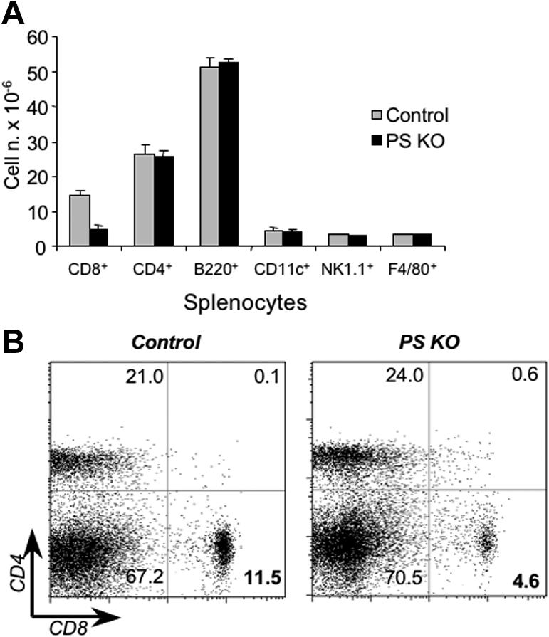
Figure 5.

Phenotypic characterization of PS KO animals. Splenocytes (A) or lymph node cells (B) from control (left panels) or PS KO animals (right panels) were staining with fluorescent-labeled mAb against CD4, CD8, CD11c, CD19, NK1.1, and F4/80. Representative of 3 animals. (B) Value in each quadrant represents the percentage of CD4 or CD8 T-cell populations inside the splenocytes of lymph node cells.
