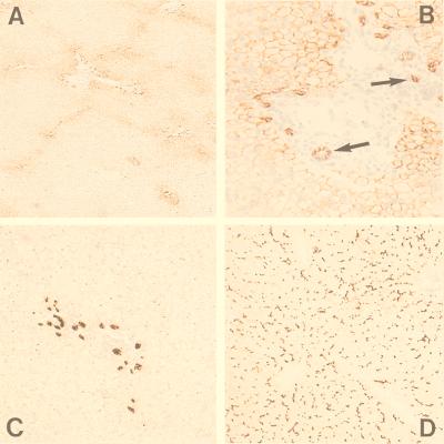
Figure 1.
Immunohistochemical staining of human liver. (A and B) Immunohistochemical detection of MRP3 in the lateral membranes of cholangiocytes (indicated by arrows) and (baso)lateral membranes of hepatocytes surrounding the portal tracts by using monoclonal antibody M3II-9. B is an enlargement of A. (C) Immunohistochemical detection of cytokeratin 7 in bile-duct epithelium cells. (D) Immunostaining with M2III-6 against MRP2, showing canalicular staining of the hepatocytes through out the entire liver lobule.

