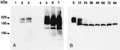
Figure 2.
Western blot analysis of MRP3 protein in parental cells and several clones of transduced 2008 and MDCKII cells. Total cell lysates (10 μg/lane for the 2008 cells and 5 μg for the MDCKII cells) were size fractionated in a 7.5% polyacrylamide gel containing 0.5% SDS. The fractionated proteins were transferred to a nitrocellulose membrane, and MRP3 was detected by incubation with monoclonal antibody M3II-9. (A) 1 = 2008; 2 = 2008/MRP3–4; 3 = 2008/MRP3–8; 4 = MDCKII; 5 = MDCKII/MRP3–17; 6 = MDCKII/MRP3–18; 7 = MDCKII/MRP3–20. (B) Time course in hours of MRP3 expression in 2008/MRP3–8 cells after treatment with tunicamycin (5 μg/ml). Ten μg of total cell lysate was used per lane.

