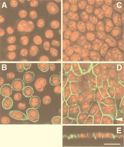
Figure 3.
Immunolocalization of MRP3 in 2008 cells and MDCKII monolayers by confocal laser scanning microscopy. MRP3 is detected by indirect immunofluorescence (green signal) with monoclonal antibody M3II-9. Nucleic acids were detected by counterstaining with propidium iodide (red signal). (A) Parental 2008 cells; (B) 2008/MRP3–8 cells; (C) parental MDCKII cells; (D) MDCKII/MRP3–17 cells; (E) vertical X/Z section of the monolayer shown in D. The arrow in D indicates the position where the X/Z section was made. (Bar = 20 μm.)

