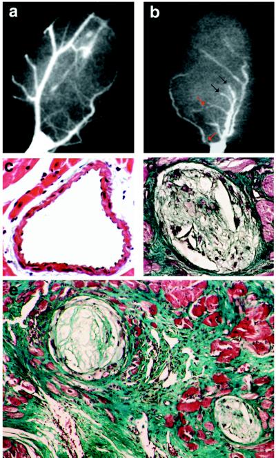
Figure 1.
Coronary atherosclerosis and myocardial postinfarction scars in hypercholesterolemic mice. (a and b) Coronary angiograms from a wild-type mouse (a) and a 7-month-old E0 × LDLR0 mouse (b). Red arrows indicate severe coronary stenosis, and black arrows indicate total occlusion. (c–e) Histological analysis of coronary arteries from wild-type (c) and E0 × LDLR0 mice (d and e). Atherosclerotic plaques are evident in the latter. In e, a myocardial scar surrounds the occluded atherosclerotic artery. [Masson’s trichrome stain, original magnification = ×200 (c and d) and ×100 (e).]

