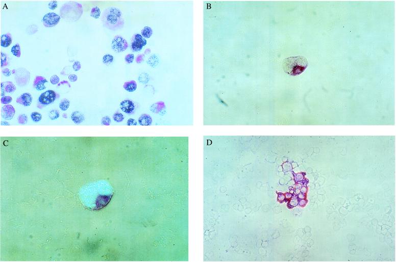
Figure 1.
Double staining of (A) p53-positive/CK18-positive PancTu pancreas cancer cells (magnification: ×85), (B) a dividing p53-positive/CK18-positive gastric cancer cell (magnification: ×136), (C) same cell as shown in B, epi-polarization microscopy (magnification: ×170), and (D) p53-negative/CK18-positive lung cancer cells (magnification: ×85). All specimens were stained with mAb CK2 against CK18 (red cytoplasmatic stain) and mAb Pab 1801 against p53 protein (blue nuclear stain).

