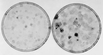
Figure 6.
The appearance of dense foci within light foci in an extended 1° assay of the original uncloned SA′ cells. The original uncloned SA′ cells were thawed, and 105 cells were cultured for 3 days in 10% CS before subculture of 105 cells for a 1° assay in 2% CS. A culture was fixed and stained at 14 days, when light foci were apparent, with occasional more densely stained cells in the center of a few foci. Other cultures were fixed and stained at 21 days when there was grossly visible evidence that denser foci were forming within the light ones. (Left) 14 days; (Right) 21 days.

