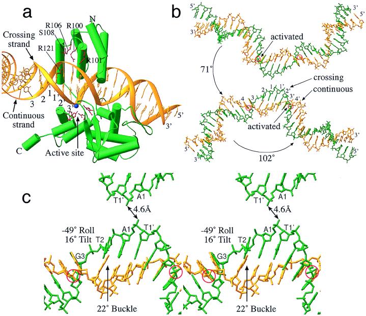
Figure 3.
The Cre R173K/loxS complex and bending of the loxS DNA. (a) A “cleaving” Cre subunit bound to a loxS half-site. Active-site side chains and side chains contacting the continuous DNA strand are drawn in red. The scissile phosphate in the active site is represented by a blue sphere. The view is from the center of the synapse. (b) Synapsed loxS sites, with scissile phosphates circled and the bound recombinases omitted. Phosphates activated for cleavage are indicated. Angles are between the 13-bp inverted repeat segments of the DNA arms (see text). (c) Close-up stereo representation of the loxS bend shown in b. The view directions of b and c are the same as in Fig. 2.

