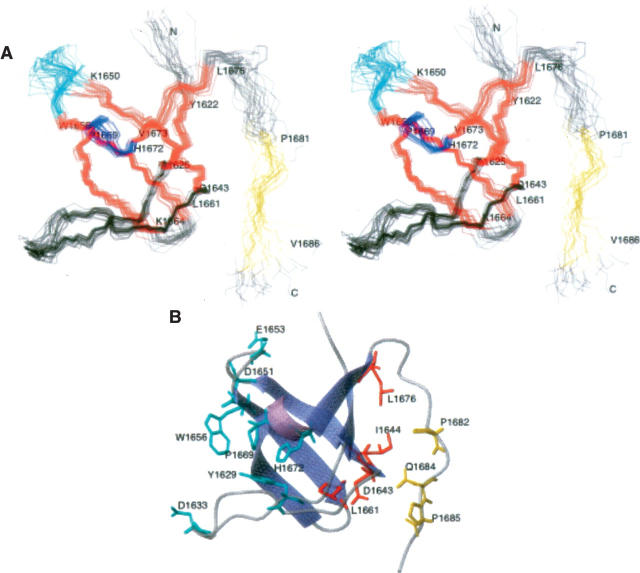
Figure 4.
The DdM7 SH3 domain interacts with an adjacent “PxxP” motif through an unexpected surface. (A) A stereoview of the 20 structures with no NOE violations above 0.5 Å are displayed with β-strands in red and the helix in blue. The region that has NOE interactions with the SH3 domain is highlighted in yellow, whereas the RT and n-Src loops are displayed in black and cyan, respectively. (B) Representative ribbon diagram of the residues A1620–H1687. The side chain atoms of residues in the region previously identified for binding “PxxP” motifs are included in cyan. Interacting residues of the SH3 domain or proline-rich region are displayed in red or yellow, respectively.

