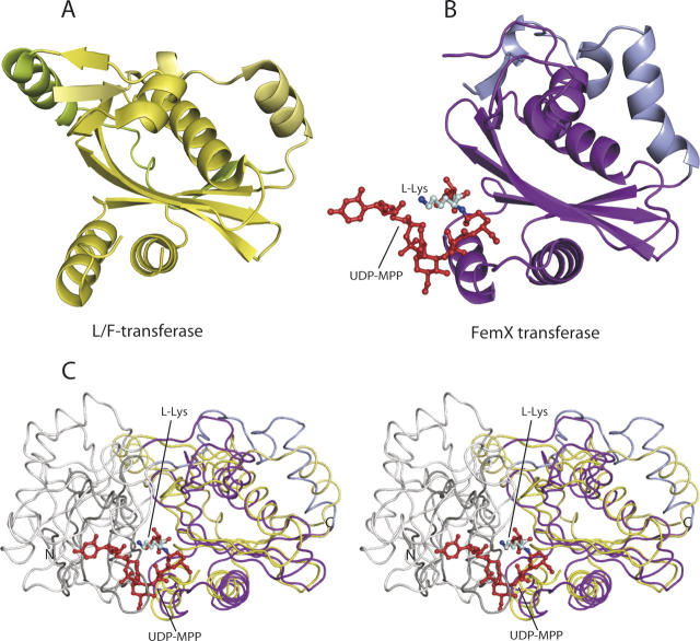
Figure 4.
Structural comparison of the L/F-transferase and the FemX transferase. The conserved GNAT superfamily fold regions are shown in yellow and purple-blue color, respectively, in the structure of the C-terminal domain of L/F-transferase (A) and FemX transferase (B), belonging to the peptidyltransferase enzymes. The UDP-MPP bound in the FemX transferase structure is colored red and depicted as a ball-and-stick model. The L-Lys of UDP-MPP is shown in pale cyan. (C) Superposition of the overall backbone structures of L/F-transferase and FemX transferase (stereo view). The structures are indicated by a main-chain, with the N-terminal domains of L/F-transferase and FemX colored dark gray and gray, respectively. The C-terminal domains of L/F-transferase and FemX are colored as in A and B, respectively.

