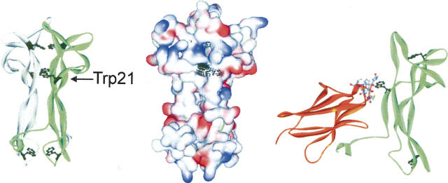
Figure 7.
Structure of NGF and its complex with the extracellular domain 5 of the TrkA receptor (PDB file 1www.pdb [Wiesmann et al. 1999]). The panels show a scheme of dimeric NGF with the Trp residues marked (black, left), the surface of dimeric NGF with the surface of Trp residues marked (black, middle), and the complex of a monomeric subunit of NGF with TrkA (right), where Trp21 of NGF and the surrounding residues of domain 5 of TrkA (8 Å distance) are shown in detail.

