
Figure 2.
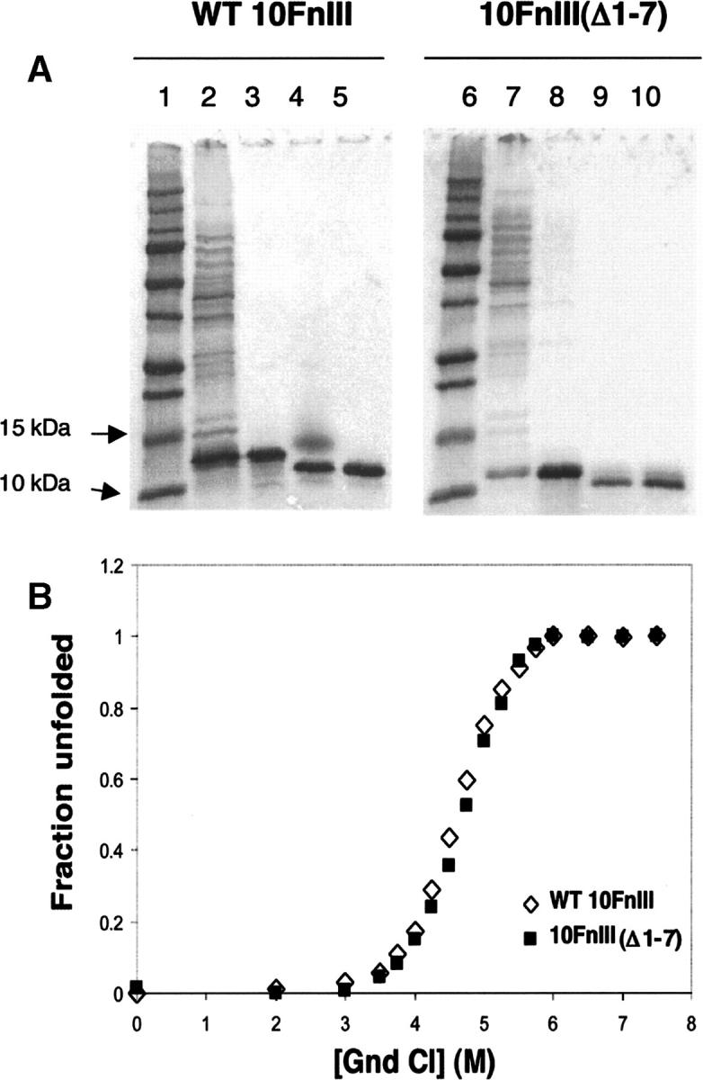
Purification and stability of WT 10FnIII compared to 10FnIII(Δ1–7). (A) Purification of WT 10FnIII (lanes 1–5) and 10FnIII(Δ1–7) (lanes 6–10). (Lanes 1,6) Protein standard. (Lanes 2,7) Cleared lysate. (Lanes 3,8) HisTrap column purification. (Lanes 4,9) Factor Xa cleavage of His6-tag. (Lanes 5,10) Gel filtration purification. (B) Guanidinium chloride denaturation of WT 10FnIII (open diamonds) and 10FnIII(Δ1–7) (closed squares) monitored by Trp fluorescence.