
Figure 1.
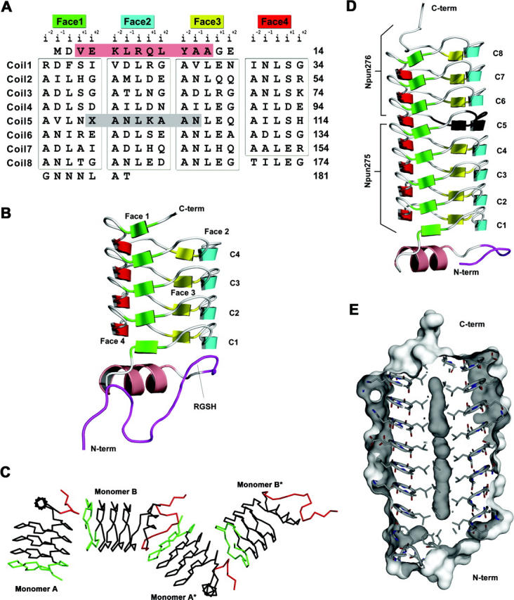
Primary and three-dimensional structures of Np275 and Np275/276. (A) Structure-based sequence alignment of Np275/276. The 31 pentapeptide repeats that are in the RHQBH conformation (boxed) are organized by their location on the faces of the quadrilateral. (Salmon) The single α-helix, (dark gray) the eight residues encoded by the DNA located between Np275 and Np276. For Np275/276 the stop codon (X) was mutated to Gln. (B) Ribbon representation of Np275. (Maroon) Residues originating from the N-terminal cleavable 6×-His-tag. (C) Np275 crystal contacts between monomers involving the N-terminal 6×-His-tag (red) and the exposed C-terminal coil (green). (D) Ribbon representation of Np275/276. (Black) The “stitching” residues between Np275 and Np276, (maroon) residues originating from the N-terminal cleavable 6×-His-tag. (E) Connolly surface of Np275/276.