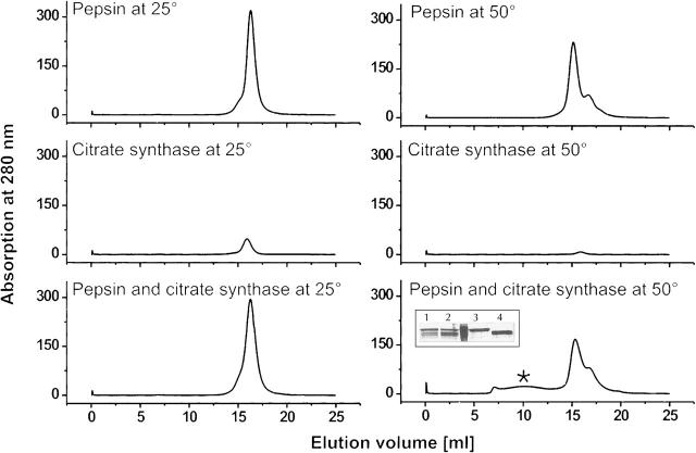
Figure 5.
Complex formation of pepsin with denatured citrate synthase. UV-profiles of gel-size exclusion chromatography runs. The asterisk (*) (bottom, right) indicates the additional peak occurring when pepsin and citrate synthase are incubated together at 50°C. The inset shows SDS-PAGE analysis of the run with the mixture of citrate synthase and pepsin. Loaded samples are: fraction at 10 mL elution (1), fraction at 15 mL elution volume (2), references citrate synthase (3), and pepsin (4).

