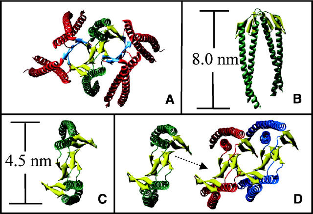
Figure 7.
Ribbon diagrams of (A) α/β PFD from M. thermoautotrophicum, based on the crystal structure (Siegert et al. 2000) showing the α PFD helices in green, the β PFD helices in red, and the double β-barrel interface in orange; (B, C) approximate dimensions of an α PFD dimer from M. thermoautotrophicum; (D) potential assembly of the γ PFD filament (top view), based on homology modeling, showing the stacking of β-strands in the same manner as for the α/β PFD.

