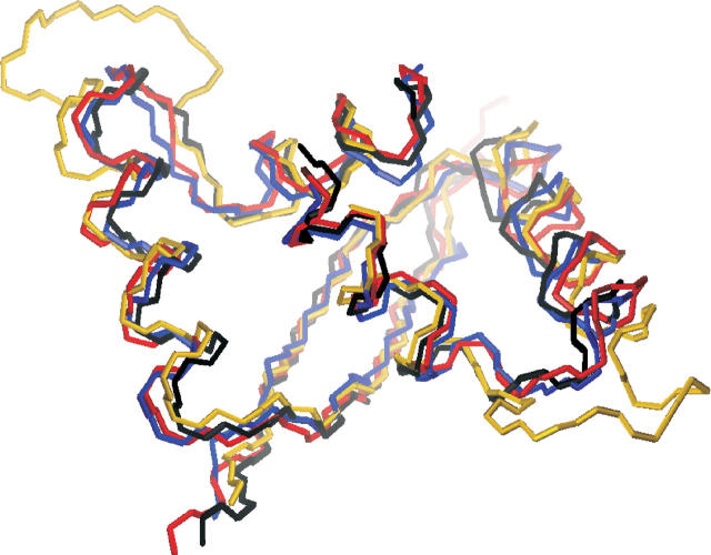
Figure 4.
Structure comparison of the ParD with the RHH proteins Arc, CopG, and MetJ. The backbone of ParD was superimposed on the backbone atoms of other RHH domains using the program MOLMOL. The aligned residues can be seen in Table 2. A view from the top of the backbone traces of ParD (blue), CopG (black), Arc (red), and MetJ (yellow) is shown.

