
Figure 1.
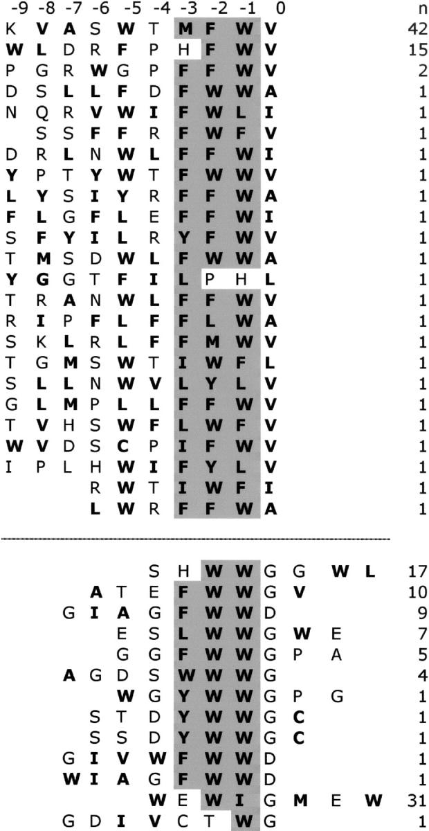
Peptide ligands for HtrA2-PDZ selected from phage-displayed libraries. Sequences selected from libraries fused to the C or N terminus of a phage coat protein are shown above or below the dashed line, respectively. Hydrophobic residues are shown in bold text, and the conserved, hydrophobic tripeptide common to the C- and N-terminal ligands is shaded gray. The number of times each sequence occurred (n) is shown to the right. Positions in the peptide ligand are numbered according to the convention for C-terminal PDZ domain ligands; residues from the C terminus to the N terminus are designated 0, −1, −2, ....