Figure 3.
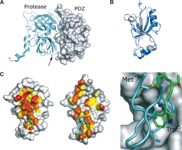
Comparison of HtrA2-PDZ bound to a C-terminal peptide and from the structure of the full-length HtrA2/Omi protein. (A) The crystal structure of full-length HtrA2/Omi (PDB code 1LCY) (Li et al. 2002). (Gray surface) The PDZ domain; (cyan schematic) the protease. (Arrow) The position of the β11–β12 region of the protease that contacts the PDZ domain. (B) Overlay of the HtrA2/Omi PDZ domain bound to a C-terminal peptide (blue) and from the structure of the full-length HtrA2/Omi protein (gray). (C) Atoms forming the binding surface for the peptide (left) or the protease (middle) are colored on the PDZ domain according to their relative distance from their respective binding partner: (red) ≤3.5 Å; (orange) 3.6–4.0 Å; (yellow) 4.1–4.5 Å. Although the details of binding are different (right), the β11–β12 region (cyan) of the protease domain contacts the same region as Trp−5 and Met−3 of the C-terminal peptide ligand (green).

