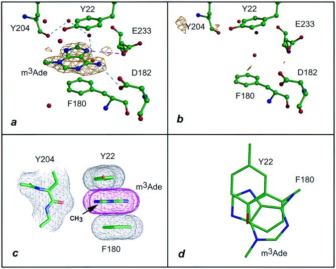
Figure 1.
Interaction of VP39 with m3Ade. (a) The 1.83-Å difference electron density of VP39 crystal soaked in 10 mM m3Ade contoured at the 2.5σ level (see Table 1 and Materials and Methods). Dashed lines represent hydrogen bonds (cyan) and salt links (magenta). (b) Equivalent to a but soaked in 10 mM adenine and with a 1.92-Å difference Fourier (Table 1). (c) Stacking of the adeninium ring of m3Ade with the aromatic side chains of Tyr 22 and Phe 180, projected parallel to the base ring. The atomic surface is shown in mesh representation. The (N)3-methyl group lies 6 Å from the peptide carbonyl oxygen of Tyr 204. (d) Same as c but projected perpendicular to the base ring with surface mesh and peptide backbone omitted for clarity.

