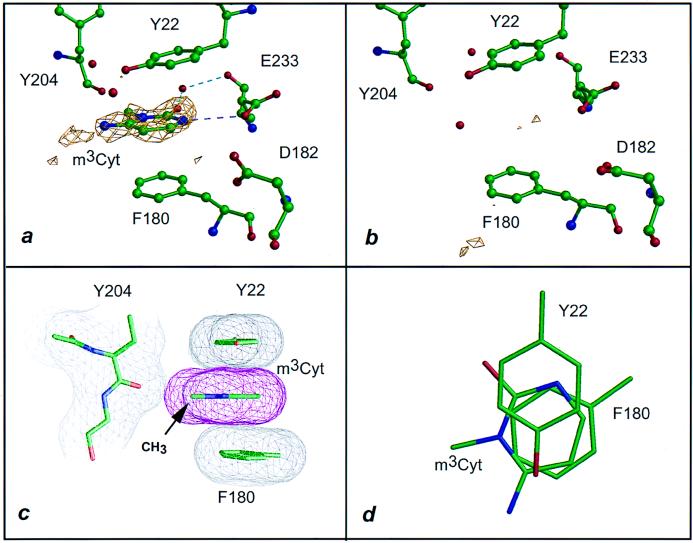
Figure 2.
Interaction of VP39 with m3Cyt. Dashed lines are colored as in Fig. 1. (a) The 1.86-Å difference electron density (2.5σ level) for VP39 crystal soaked in 10 mM m3Cyt (see Table 1 and Materials and Methods). (b) Identical to a but soaked in 10 mM Cyt (Table 1). (c) Stacking of cytosinium ring patterned after Fig. 1c. The (N)1-methyl group lies within 3.3 Å of the carbonyl oxygen of Tyr 204. (d) Same as c but fashioned after Fig. 1d.

